| dc.contributor.author | Sevinç, Özdemir | |
| dc.contributor.author | İs, Merih | |
| dc.contributor.author | Barut,Çağatay | |
| dc.contributor.author | Erdoğan, Alirıza | |
| dc.date.accessioned | 2020-12-19T19:58:56Z | |
| dc.date.available | 2020-12-19T19:58:56Z | |
| dc.date.issued | 2014 | |
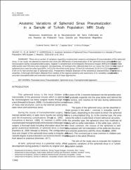
| dc.identifier.citation | Sevic, O., Is, M., Barut, C., Erdogan, A., (2014).Anatomic Variations of Sphenoid Sinus Pneumatization in a Sample of Turkish Population: MRI Study.International Journal of Morphology, 32(4), 1140-1143.https://doi.org/10.4067/S0717-95022014000400003 | |
| dc.identifier.issn | 0717-9502 | |
| dc.identifier.issn | 0717-9367 | |
| dc.identifier.uri | https://doi.org/10.4067/S0717-95022014000400003 | |
| dc.identifier.uri | https://hdl.handle.net/11436/3022 | |
| dc.description | WOS: 000348581400003 | en_US |
| dc.description.abstract | There are a number of variations regarding morphometric anatomy and degree of pneumatization of the sphenoid sinus. in our study, we planned to examine and show the differences of pneumatization of the sphenoid sinus particularly to guide the neurosurgeon during transsphenoidal surgery. Sagittal T1-weighed spin-echo Magnetic Resonance Images (MRIs) of 616 adult individuals (406 women and 210 men) were analyzed, retrospectively. According to the collected data from our study, the most common type of the sphenoid sinus was the sellar type (83%; n=511) for the whole study group. of the 616 individuals 16.6% (n=102) had presellar type and 0.5% (n=3) had conchal type of sphenoid sinus. Preoperative detailed detection of the anatomical characteristics of sphenoid sinus is essential. A thorough information obtained from studies of the regional anatomy and awareness of its variability can provide a safe and accurate transsphenoidal and extended endoscopic skull base approaches. | en_US |
| dc.language.iso | eng | en_US |
| dc.publisher | Soc Chilena Anatomia | en_US |
| dc.rights | info:eu-repo/semantics/openAccess | en_US |
| dc.subject | Paranasal sinus | en_US |
| dc.subject | Pneumatization | en_US |
| dc.subject | Sphenoid sinus | en_US |
| dc.subject | Transsphenoidal approach | en_US |
| dc.title | Anatomic variations of sphenoid sinus pneumatization in a sample of Turkish Population: MRI study | en_US |
| dc.type | article | en_US |
| dc.contributor.department | RTEÜ, Tıp Fakültesi, Temel Tıp Bilimleri Bölümü | en_US |
| dc.contributor.institutionauthor | Sevinç, Özdemir | |
| dc.identifier.doi | 10.4067/S0717-95022014000400003 | |
| dc.identifier.volume | 32 | en_US |
| dc.identifier.issue | 4 | en_US |
| dc.identifier.startpage | 1140 | en_US |
| dc.identifier.endpage | 1143 | en_US |
| dc.ri.edit | oa | en_US |
| dc.relation.journal | International Journal of Morphology | en_US |
| dc.relation.publicationcategory | Makale - Uluslararası Hakemli Dergi - Kurum Öğretim Elemanı | en_US |