| dc.contributor.author | Yılmaz, Rukiye | |
| dc.contributor.author | Bedir, Recep | |
| dc.date.accessioned | 2022-07-29T06:56:26Z | |
| dc.date.available | 2022-07-29T06:56:26Z | |
| dc.date.issued | 2021 | en_US |
| dc.identifier.citation | Yilmaz, R., & Bedİr, R. (2021). Pulmonary Acantholytic Squamous Cell Carcinoma Mimicking Lepidic Pattern Adenocarcinoma. Pulmonary Acantholytic Squamous Cell Carcinoma Mimicking Lepidic Pattern Adenocarcinoma. Turk patoloji dergisi, 37(1), 89–91. https://doi.org/10.5146/tjpath.2019.01486 | en_US |
| dc.identifier.issn | 1018-5615 | |
| dc.identifier.issn | 1309-5730 | |
| dc.identifier.uri | https://doi.org/10.5146/tjpath.2019.01486 | |
| dc.identifier.uri | https://hdl.handle.net/11436/6286 | |
| dc.description.abstract | Dear Editor,
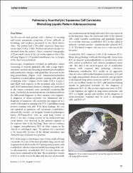
An 84-year-old male patient with a history of smoking and recent pneumonia symptoms of fever, difficulty in breathing, and weakness presented to the chest disease clinic. The patient had a left-sided suspicious lung mass on his chest X-Ray (CXR). Written informed consent was obtained from the patient. Chest computed tomography (CT) revealed a mass of 3x2 cm on the superior lobe of the left lung (Figure 1). CT-guided transthoracic tru-cut biopsy of the chest was performed. | en_US |
| dc.language.iso | eng | en_US |
| dc.publisher | Fedaration Turkish Pathology Soc. | en_US |
| dc.rights | info:eu-repo/semantics/openAccess | en_US |
| dc.title | Pulmonary acantholytic squamous cell carcinoma mimicking lepidic pattern adenocarcinoma | en_US |
| dc.type | letter | en_US |
| dc.contributor.department | RTEÜ, Tıp Fakültesi, Cerrahi Tıp Bilimleri Bölümü | en_US |
| dc.contributor.institutionauthor | Yılmaz, Rukiye | |
| dc.contributor.institutionauthor | Bedir, Recep | |
| dc.identifier.doi | 10.5146/tjpath.2019.01486 | en_US |
| dc.identifier.volume | 37 | en_US |
| dc.identifier.issue | 1 | en_US |
| dc.identifier.startpage | 89 | en_US |
| dc.identifier.endpage | 91 | en_US |
| dc.relation.journal | Turkish Journal of Pathology | en_US |
| dc.relation.publicationcategory | Diğer | en_US |